Treatment of thigh bone fractures with four heads, hand and shoulder and lower bones and upper
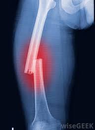
Coalescence of fractures The treatment of fractures and docking with different ways, depending on the degree of elevation or runny blood to Bone, and the bone around, and on the degree and location of the injury, and the age of the patient, and finally on Method of treatment used. Broke the bone always mean poor blood rod to the bone, and all the blood disorder and Rhode impedes Bone healing process and his tackle, while the vigor and vitality of blood vessels is one of the Are essential in the ossification process, so heal shin bone rectangular be While weak heal immediately basal parties bone, the area responsible for the divisions (Cellular bone) because they are rich vibrant, and not through the periosteum, which coats the bone Through the joint, ligaments, tendons close it. The Albanian cell to maximize (docking) formed on the surface of the broken bone, and the bone Soft (sponge) have covered the surface is very large, while the bone hoarder "Extreme coherent" be limited exclusively in the lining of the spinal cord channel Central, and moreover Valazm soft (sponge) is best fed with blood, Therefore, the adhesion is faster than the bone-ROM (Compact). Stages of fusion breakage This should be taken into account when assessing the treatment period, the total blood flow torrent In one of the broken fragments greatness femoral neck or wrist greatly prolongs the period of bone adhesion. Coalescence fractures in X-rays The congestion is caused by the bloody break. During the twelve days is subject to congestion For a particular system. In this period, the movement of the bone fragments can be observed, while appearing in X-sharp cracks or simple fractions and after the process of absorption of tension fabric shows Connective Alaltahami in the case. The initial docking of bone that occurs in the bone tissue contains fragments of Hydroxy Ibnat, and appears in the X-chips such as wool (wool yarn). The parties to the bone Not sharp, and appear on the form of fog (in young people, this show within a few days, and in the Adults from three to four weeks). Biology (vital), the docking marrow appears on the X-like spongy bone, Clearly seen on the bone rectangle. Adhesions and bony take place within three to six months, however, the notch Fractional could occasionally seen after a year and a half, such as those after the duration, or when it happens Total docking bone again, the place where the fracture completely disappear. The information listed are only guidelines for the importance of, and the legal task it requires Preparation and special care to determine the injury, and the date of occurrence and offal. Fusion of skeletal fractures and cured in adults requires anchor calendar limited Time periods (and preventive treatment) as follows: Alazmamdh type of installation Shoulder The upper part of the radius bone The lower part of the radius bone Body of radius Crank (one bone) (Crossbones) The neck of the femur Femoral trochanter area The lower part of the thigh The upper part of the leg Shin The lower part of the leg Ankle area 3 - 5 weeks 4-5 weeks 4-6 weeks 8-10 weeks 6-8 weeks 10-12 weeks 12-16 weeks 8-10 weeks 10-12 weeks 8-10 weeks 10-16 weeks 10-16 weeks... Physiological fusion fractures Congestion of the blood vessels, which Ttglt during 6-8 hours injury arises. And thrombosis associated with a broken bone operates as an intermediary for the proliferation of new cells in the cells Connective tissue between the bone fragments and to form the nucleus of the maker of the new bone. In the early days Following the break, there is the connective tissue between the bone fragments, then quickly show tissue Cicatricial Or granular. In the next stage, and on the newly formed blood vessels, Precipitate Constituent of bone material (a bag skeleton) and produces the so-called bone coherent, and means This phase fragments touch Article initial fusion (Callus) broken bones Such as clinical skeletal Union, which requires several periods, based on the type and size of the bone. In the next phase of treatment, the Albanian cell absorption of bone leads to the replacement Coherent bone bone Mussafah which relates to the composition of lines and load pressure forces the bone ( Afford it). The method described in the docking marrow take place under certain conditions. For Example: the weakness of the installation process (splinting) and complications of Osteopathic installation not only be Seriously annoying, but thus provide adhesion and bone formation detailed false (false - contact Bony placebo). This, and the pollution and inflammation and infection of the fractured bone can disrupt his tackle for a month or Years, so Shell docking marrow contaminated process takes longer. The treatment of fractures requires taking into account the conditions of several chemical and biological tissue, and mechanical ... Although fractures calendar of important things for a sports person to be able to practice his sport. Coalescence fractures disorders Atrophy of skeletal Statistics class, especially in cases of shrapnel fractional surface, be A natural evolution in Alaltahami bone to break. The Sodic exhibitors from serious complications Which affects the mobility and strength of the foot. In the first phase there are complications on the base of Food effect of the nerves in the form of swelling and redness of the skin phenomenon. The X-rays show atrophy skeleton Patchy (only). If the development of the condition continues The symptoms persist, it could get inflation bony tissue Hyper plastic bone)) . Delay or absence of significant adhesions in the complex process of docking skeletal complications. Late adhesions take its place with the continuity of Osteopathic installation (Immobilization) appropriate and proper re-installation of the parties and the broken fragments of bone, and During the X-spacing ends parties bone fracture and a widening fissure fractional show. Late adhesions can be docking in the case of the decay of the bone and his over-zealous, but this The phenomenon of the decomposition of bone and Triqgaha accompany these cases. Can false joint between the parties to the fracture configuration, and this is in the case of decay or not Appropriate reconstructive methods (internal and external reasons). Ray image shows a lack of docking Marrow, rotated gradually, forming a typical bone fracture parties. And the closure of channels Medullary bone marrow and widening fissure, and be detailed false instead of docking Bone and roughly the bones of his relationship thigh and upper arm bones of the hand does not exceed And foot. The emergence of a clear increase in packets of tartar to the parties to the broken bone and gradual closure Channels medullary bone cortex and weak interaction and the widening fissure bone fracture zone All of which give a clear diagnosis for late adhesion false detailed composition process. Unfortunately, it is very natural to be delayed for a change in the course of the case, which reached To injury. On this diagnosis in cases of difficult situations need to be applied imaging And X-rays glass to clarify the situation. Applied Imaging despite foggy and installation of barrier marrow, they illustrate well how and expansion The ends of the broken bone rigid (Sclerosis). Pictures magnifying able to give details More pronounced for the installation of the bone, and the restoration of the bone on the other hand. As a result of individual injuries, Accompanied by multiple fractures, can be accompanied by muscle calcification after infection, and in varying Different because of the rupture of the cortex bone fragments fractures, and muscle brachial gibbet of the most Muscles prone to calcification.... Calcification of bone can be seen above the extrusion coronal (calcification appear hazy and then Take the form Osvenjia). As can be seen above the muscle calcification trigonometric and muscle widening Inside the femoral. Tissue calcification and encircles the full shift (caught) connective tissues Overlapping parts of the muscle. Such calcified muscles appear in radiology through 3-5 Weeks, and increase to 6-8 months after infection. The access to these complications The barriers are very important, because it reveals early on calcified muscle. Chronic fractures These cases are caused by continuous endurance excess bone, working on excessive resistance Physiological, and ease of occurrence minute, especially sports-related (fractures injuries Debilitating or tired from areas prone to increase endurance, such as second and third paragraphs And fourth paragraphs of cervical, and foot and thigh, with the exception of the inner surface of the femur The trachea and heels). In many cases, fractures can not can not see the incision in X, and this explains the rich fusion and interaction in the bone cortex (periosteum), and if you take Image rays early, it can be seen clearly notch. Detailed false Pseudoarthrosis This consists in the place of the fracture, which was due to some reasons the parties do not coalesce with fracture Each other. After two or three months in place of the fracture, and rather than be docking Bone, cartilage tissue consists of (brittle) and this will be detailed and placebo. In such cases, Valjrahh is the only treatment, and this is made up in the following points: 1. Remove the configuration cartilage 2. revive the edges of the parties fractures and bone using pruning. 3. Action landing install metal (osteosinthesis). Pruning is taking bone marrow crust of the leg area of the young upper area Bone (epiphysis). And digging long channel between naughty and pen bone fracture is placed in the stream Channel, and this usually proves Palmthbtat cuneiform metal. User injured and edger placed in broken install Gypsum for 3-6 months. If an event in Milan Another axis of the bone and beyond any growth after surgical intervention, there is no objection from another surgical intervention To evaluate the warp winning, and an amendment bone (osteotomy). And here takes into account the length of Lists The infected patient, out of fear of what the Palace. Many scientists believe that the reasons for the false joint occurrence lies in hormonal factors affect the Metabolism of the components of the bone on top of calcium, as well as the softer bone Acquired in some people. Sudeck's atrophy of bone: It is the demise of mineral salts from the infected bone opposed, and loss of elasticity of the soft tissue Surrounding the fracture, as a result of poor blood circulation, especially in the articular region prone To infection. He spoke these symptoms in most cases in the joints of the lower limbs and the attendant Status radiating pain in the affected area, and poor circulation as a result of tight blood (Vasoconstriction) happens after the paralysis of the puppet aneurysm (vasodilation) In the case First blood vessels shrunken (Mnaqbdh) and cellular poor nutrition while in case The second blood Valawaah expansive and slow speed of blood flow and the recent result is One. And that the deficit in the cellular nutrition results are: 1. demise of bone mineral salts. 2. The loss of elasticity of the soft tissues. 3. Muscular Dystrophy. These variables are a serious impediment occur if preventive therapeutic methods are not followed to prevent Evolution. We can consider the offer Sodic paralysis of the blood vessels and pass this projector in three stages Succession: The first stage: the stage of pain and swelling hinge called the acute phase. Second stage: the stage of racial decomposition of metals and leachability of the bone. Third stage: the stage of continuity and decay metal salt of the bone (change structure Bone). The clinical picture of the situation: Articulated radioactive pain in the affected area, swelling and swelling, Redness of the skin, chilled articular region, and infect a cold sweat and movement painful articular And limited. The surrounding soft tissue atrophy in the case of the occurrence of articular stiffness. Therapeutic intervention: The therapeutic intervention varies from one person to another due to varying degrees of injury among persons Therapeutic accurate and interference in stages and its application. And it must take the following steps: - Lack of detailed Qbarp (violent) motor. - Hinge installation does not give satisfactory results. - Kinetic applied by the processor should be under the limits of pain, because this can Cause atrophy. - The application of therapeutic support and active movements that depend on the patient. - Repeated several times a day therapeutic movements. - Raise the physical ability when injured. - The use of water means (water treatment) and electric therapy in the treatment of the case.
ليست هناك تعليقات:
إرسال تعليق